

An Inflammatory Response
The body reacts to injury, including brain injury, with a predictable inflammatory response. Swelling (edema) is a manifestation of that response and is the result of increased blood flow which brings white blood cells to the injured site, and the movement of other fluids, to help heal the injury. Edema is a sign of an underlying problem, but not the disease/injury itself. Brain swelling (cerebral edema), which can last up to 5 days post-trauma, can be in specific locations within the brain, or over widespread regions of the brain, and can be a direct or indirect response to trauma.
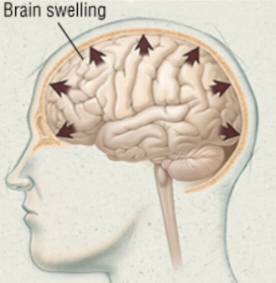
Unlike swelling in most other parts of the body, cerebral edema can cause serious injury and even death if not treated quickly. It can also be difficult to diagnose without proper testing and a thorough examination. Make no mistake, brain swelling following trauma is a very serious condition.
Intracranial Pressure
Management of cerebral edema focuses on controlling intracranial pressure. Intracranial pressure (ICP) is a measurement of the combined pressure inside the skull of both brain tissue and cerebrospinal fluid. Following trauma, increasing ICP in this enclosed environment is a definitive sign of brain swelling.
Realize that when the brain swells there’s no place for it to go. It’s encased inside the rigid confines of the skull which does not expand. Since there’s no room for expansion, swelling causes the brain to become compressed. This occurs because the brain is soft, has the consistency of gelatin, and therefore compresses easily. This compression squeezes the blood vessels inside the brain restricting blood flow, thereby depriving brain tissue of the blood oxygen and blood sugar (glucose) it needs to function and survive. Swelling can also block cerebral spinal fluid, which circulates through the brain and into the spinal cord, from exiting the brain which can make the swelling worse and ICP even higher. If unchecked, swelling can crush brain tissue, shift brain structures from their normal locations, contribute to hydrocephalus (excess water on the brain), and can even cause “brain herniation” where a part of the brain is squeezed across and into other structures, such as the brain stem. Brain herniation is almost always fatal.
ICP can be measured. Measuring requires surgical placement by a neurosurgeon of a small pressure-sensitive probe. The probe is inserted through a small hole drilled in the skull and placed a short distance into the brain. This allows for ongoing, continuous monitoring of ICP.
Signs and Symptoms
Signs and symptoms of cerebral edema include:
- headache
- dizziness
- nausea
- lack of coordination
- numbness
In more severe cases of cerebral edema, you may experience symptoms including:
- mood changes
- memory loss
- difficulty speaking
- incontinence
- changes in consciousness
- seizures
- weakness
- double vision
Therapeutic Measures
Those who are experiencing brain swelling should be hospitalized and closely monitored during the first 48 to 72 hours after injury. Various diagnostic protocols begin with a CT scan or MRI, with an MRI being far more diagnostic. Once a brain swelling diagnosis is made, efforts are undertaken to relieve the pressure within the skull cavity caused by the swelling until the swelling eventually subsides. These include:
- keeping the hospital bed elevated 30 to 45 degrees. When the head is flat it can increase pressure in the brain.
- maintaining normal body temperature with antipyretics (fever reducers)
- maintaining a calm environment with low lighting to avoid agitation.
- monitoring fluid and electrolyte levels
- administering anticonvulsants for seizure prevention
- prescribing pain relievers to increase comfort
- draining cerebral spinal fluid by inserting a catheter (shunt) into the brain ventricles
- administrating neuromuscular blockades (medications to induce paralysis), and
- offering hyperosmolar therapy which allows cerebrospinal fluid to move/flow from inside the cranial space (skull) reducing ICP.
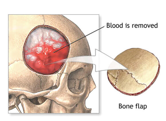
In extreme and emergency situations, a surgery may be necessary to lower the pressure. If the elevated ICP is the result of a brain bleed (hematoma), an aspiration of the pooled blood may be necessary. Aspiration involves drilling a small hole in the skull and using a needle to evacuate the blood. A more invasive procedure is a craniotomy (partial removal of a portion of the skull). In this procedure blood evacuation is made easier. This is obviously a major surgical event and is typically used only when a hematoma is large, when more room inside the skull must be created for the swelling brain to expand, or when the swelling starts to compress the brain stem where critical functions are performed, like breathing, swallowing, heart rate and blood pressure maintenance.